
You wore a full upper denture for years. It kept getting looser. You finally saw a dentist about implants and heard the same thing every clinic told you: "There's not enough bone." That verdict feels final — but for patients with severe maxillary atrophy, it often isn't. Beijing's top-tier stomatology hospitals run dedicated programs for exactly this category of case, with surgical volumes and specialist access that are genuinely difficult to match in the West.
Quick Decision Snapshot
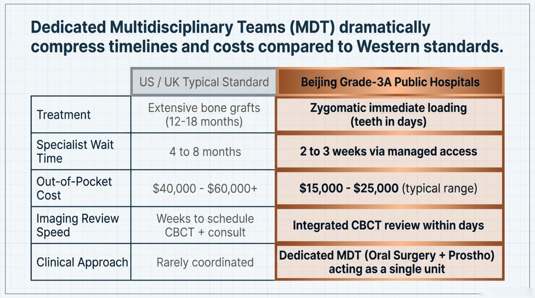
| Key Factor | US / UK Typical Standard | Beijing Grade-3A Public Hospitals |
|---|---|---|
| Treatment for Severe Atrophy | Extensive bone grafts (12–18 months) | Zygomatic implants with immediate loading — teeth in days, not years |
| Specialist Wait Time | 4 to 8 months | 2 to 3 weeks via managed access |
| Out-of-Pocket Cost | $40,000–$60,000+ | $15,000–$25,000 (typical range) |
| Imaging Review Speed | Weeks to schedule CBCT + consult | CBCT review integrated into surgical planning within days |
| Multidisciplinary Review | Rarely coordinated for implant cases | Dedicated MDT (Oral Surgery + Prosthodontics) for complex atrophy cases |
If you already have a CBCT scan or panoramic X-ray, you don't need new records to get started. Email what you have to contact@pandamedglobal.com — the team can check whether your existing imaging is enough for an initial feasibility review and tell you exactly what else, if anything, to prepare.
Why "No Bone" Doesn't Always Mean "No Implants"
When maxillary bone resorption reaches Cawood and Howell Class V or VI — the classification used in established oral surgery guidelines for extreme alveolar atrophy — the residual ridge is too thin and too shallow to anchor a standard implant. Attempting large-volume bone grafts in this state carries high morbidity: multiple surgeries, a 12-to-18-month healing window, and a meaningful risk of graft failure before a single implant is even placed.
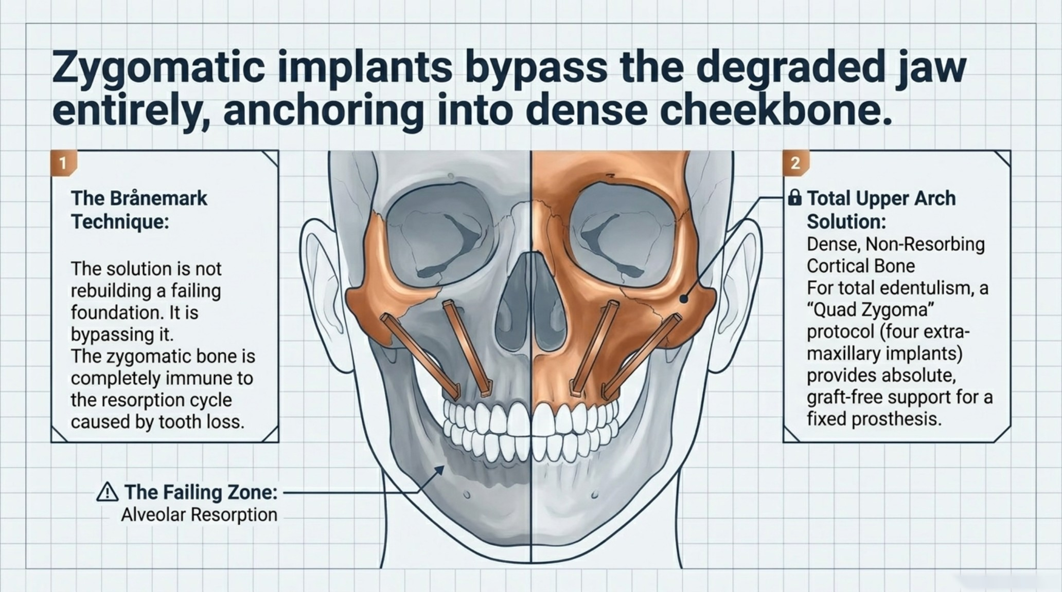
Zygomatic implants take a fundamentally different approach. Rather than rebuilding the missing jaw, they bypass it entirely. The implants anchor into the zygomatic bone — the cheekbone — which is structurally dense, well-vascularized, and does not resorb the way alveolar bone does after tooth loss. The technique was originally developed by Professor Per-Ingvar Brånemark and has been refined over decades into a well-documented solution for patients who have exhausted conventional options.
For patients with total upper arch edentulism and severe atrophy, a "Quad Zygoma" configuration — four zygomatic implants, two per side — can provide the full-arch support needed for a fixed prosthesis without any bone grafting at all.
The Immediate Loading Advantage
One of the most clinically significant features of zygomatic implants is their primary stability. Because the cheekbone is dense cortical bone, the implants achieve high insertion torque values at placement. This mechanical stability is what makes immediate loading possible in appropriate cases: a fixed provisional bridge can be attached within 24 to 48 hours after surgery.
Compare that to the conventional grafting pathway. A sinus lift or block graft requires 6 to 12 months of healing before implants can be placed. Then the implants themselves need another 3 to 6 months of osseointegration before a final prosthesis is fitted. A patient who starts that pathway today may not have a functional fixed set of teeth for 18 to 24 months — during which time continued bone resorption under a removable denture makes the situation progressively harder to manage.
For patients who have already been edentulous for years, stopping that resorption cycle matters. Immediate loading isn't just a convenience. It's a clinical reason to act sooner rather than later.
What High-Volume Beijing Centers Do Differently
Zygomatic implant surgery is widely described in the implantology literature as one of the most technically demanding procedures in the field. The implant trajectory passes through complex facial anatomy in close proximity to the maxillary sinus, the orbital floor, and surrounding soft tissue structures. Surgeon experience and case volume are not minor variables — they are central to outcomes.
In the US and most of Western Europe, zygomatic procedures are performed by a small subset of oral and maxillofacial surgeons, many of whom handle only a few cases per year. Beijing's top specialized Grade-3A stomatology hospitals operate at a fundamentally different scale:
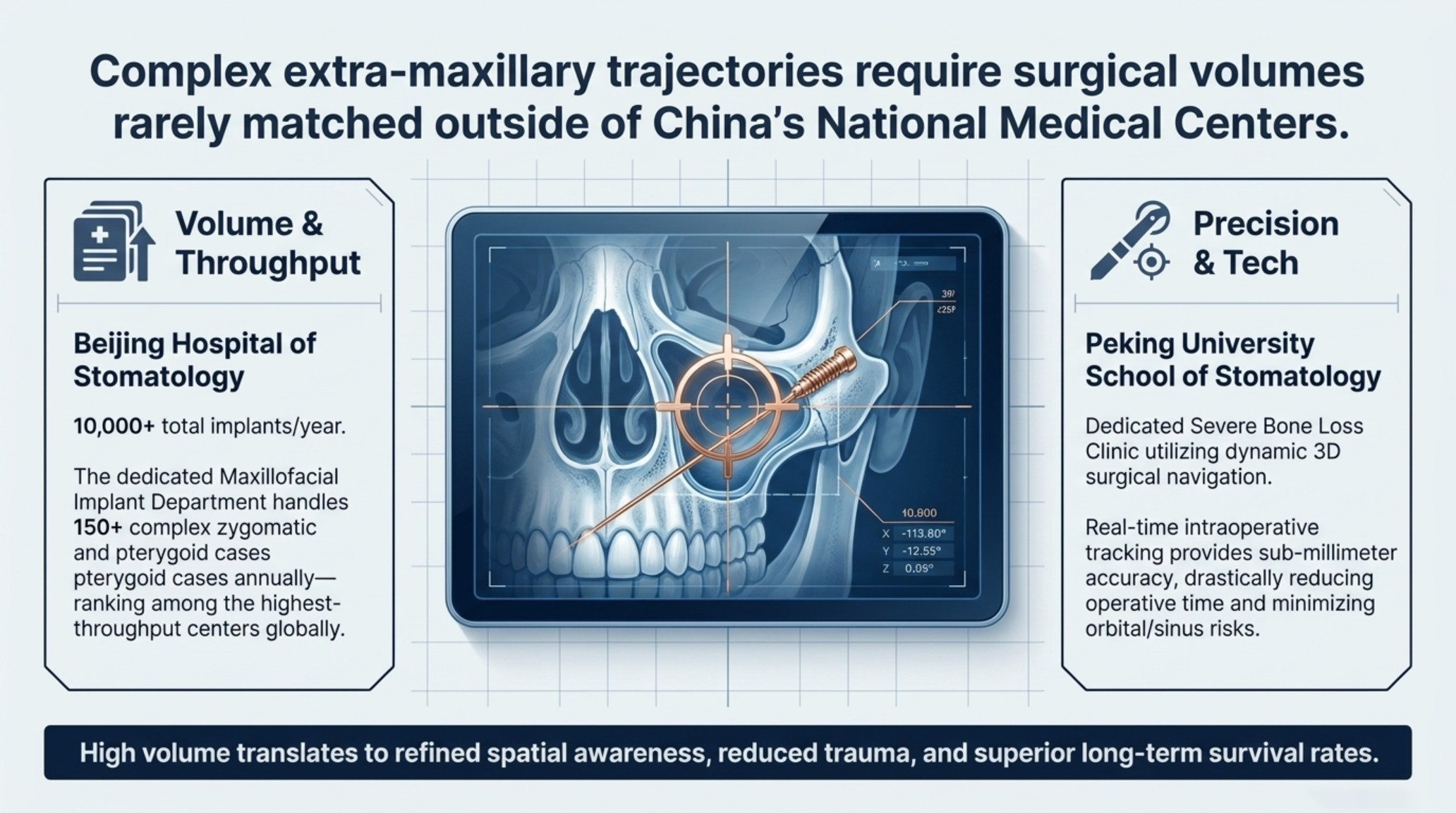
Beijing Hospital of Stomatology performs over 10,000 implant procedures annually. Its dedicated Maxillofacial Implant Department handles more than 150 complex zygomatic and pterygoid cases each year — a volume that places it among the highest-throughput centers for this procedure type globally. (Source: Department of Oral Implantology, Beijing Hospital of Stomatology, 2023 Annual Internal Review)
A second major center operates a dedicated Severe Bone Loss Implant Clinic within its oral surgery department, utilizing dynamic 3D surgical navigation systems for extra-maxillary implant placement with sub-millimeter accuracy. Real-time intraoperative navigation reduces surgical time and minimizes the risk of sinus perforation or orbital proximity complications. (Source: Clinical Pathways for Extra-Maxillary Implants, Peking University School of Stomatology, 2024 Institutional Report)
High case volume translates into something specific and measurable: surgeons develop refined spatial awareness of zygomatic anatomy, shorter operative times, reduced intraoperative trauma, and higher long-term implant survival rates. These are not abstract advantages.
What a Complex Case Actually Looks Like
A recent internal case review from one of Beijing's leading stomatology centers documented a 65-year-old patient who had worn a removable upper denture for a decade. Her maxillary bone was described as paper-thin on CBCT imaging. She had been turned away by three separate dental clinics in her home country, each citing insufficient bone for any implant approach.
Through a Beijing MDT pathway coordinating oral surgery and prosthodontics, she underwent a Quad Zygoma procedure using 3D-printed surgical guides derived from her CBCT data. The surgery was completed safely. A fixed acrylic bridge was attached within 24 hours. Within weeks, she was able to bite into solid food for the first time in years.
This type of case — full upper arch atrophy, prior rejection by multiple providers, decade-long edentulism — is not unusual at Beijing's high-volume centers. It is, in fact, the category of patient these departments are specifically structured to handle.
If your case involves prior graft failure, a history of bisphosphonate use, or complex sinus anatomy, those details matter for surgical planning. You don't need a new workup to ask the question. Email your existing scans and records to contact@pandamedglobal.com — the team can review what you have and advise on next steps.
The Checklist: Records That Help Most
You do not need brand-new records to start. Existing records are often enough for an initial feasibility check. Here is what is most useful:
CBCT Scan (highest priority): The actual 3D image files in DICOM format, exported to a shareable cloud link (Google Drive, Dropbox, or similar). A standard 2D panoramic X-ray alone is not sufficient for zygomatic surgical planning, but it is still worth sending if it's what you have.
Clinical photographs: Clear photos of the upper arch, lower arch, and occlusal view if possible.
General health panel: Zygomatic implant surgery is a medical procedure requiring anesthesia. A recent blood panel — including HbA1c for blood sugar control — is needed for pre-surgical clearance.
Medication list: Particularly important for patients taking bisphosphonates (for osteoporosis), anticoagulants, or immunosuppressants. These affect surgical planning and timing.
Prior treatment records: Any documentation from previous implant attempts, graft procedures, or specialist consultations — even if the outcome was a rejection.
Family and Logistics
For international patients, the logistics of traveling to Beijing for a multi-stage surgical procedure involve more than booking a flight. Questions about visa categories for medical travel, accommodation near the hospital, interpreter services during consultations, family accompaniment, and post-operative recovery planning are all manageable — but they require coordination.
If you need help thinking through any of these, email contact@pandamedglobal.com. The team handles translation, record formatting, appointment access, and Beijing care coordination for international patients.
Author Bio Ryan Lee is a Beijing-based medical concierge and the founder of PandaMed. With a strict compliance background, he helps international patients navigate China's top public hospitals safely. He secures direct access to Chief Physicians for complex cases.
Medical Disclaimer This information is for educational purposes only and is not medical advice. PandaMed is a care coordination service, not a medical provider. Always consult a qualified doctor for your specific condition.
References
Brånemark, P.I., et al. (2004). Zygomaticus fixture in the management of advanced atrophy of the maxilla: technique and long-term results. Scandinavian Journal of Plastic and Reconstructive Surgery and Hand Surgery, 38(2), 70–85.
Aparicio, C., et al. (2010). Zygomatic implants: indications, techniques and outcomes, and the zygomatic success code. Periodontology 2000, 52(1), 19–55.
Cawood, J.I., & Howell, R.A. (1988). A classification of the edentulous jaws. International Journal of Oral and Maxillofacial Surgery, 17(4), 232–236.
Surgical Volume Data, Department of Oral Implantology, Beijing Hospital of Stomatology (2023 Annual Internal Review). [Plain-text attribution only]
Clinical Pathways for Extra-Maxillary Implants, Peking University School of Stomatology (2024 Institutional Report). [Plain-text attribution only]
